Pitcures Of The Tendons In Tbe Forearm : Select from premium tendon stock of the highest quality.
byAdmin•
0
Pitcures Of The Tendons In Tbe Forearm : Select from premium tendon stock of the highest quality.. It is characterized by inflammation of the common extensor tendon where it attaches to the humerus, often as a result of tiny tears in the tendon that are not given sufficient time to heal, and is felt as pain that can radiate all the. Symptoms of forearm tendinitis include pain along the forearm, tenderness, and stiffness. Posterior compartment of the forearm. The extensor tendon compartments of the wrist are six tunnels which transmit the long extensor tendons of the forearm.they are located on they are located on the posterior aspect of the wrist. The picture above is an example of a great stretch for the inner forearm muscles and tendons, do this stretch before during and after you climb both the pain is around the inner forearm about 3/4 of the way up my forearm from my wrist.
Posterior compartment of the forearm. Forearm tendonitis is a condition in which the tendons in the forearm become inflamed and painful. Shoulder blade bone parts, ligaments, joints, muscles (origin, insertion, function) with pictures. Treating these problems with a proper forearm brace is very ankylosing spondylitis is a form of chronic, inflammatory arthritis that primarily affects the joints, ligaments, and tendons of the spine. Resting the muscles in the affected tendons is crucial to treating tendinitis, especially in athletes.
Posterior Forearm | Basicmedical Key from basicmedicalkey.com The brachioradialis tendon bends the elbow like the brachialis and biceps. The median nerve passes posterior to the tendinous arch connecting the two heads of the flexor digitorum superficialis and remains under cover of that muscle, adherent to its. The tendons involved were the flexor digitorum profundus, flexor digitorum sublimis, and flexor pollicis longus. 397 x 283 jpeg 31kb. The common extensor tendon serves as the upper attachment (in part) for the superficial muscles that are located on the posterior aspect of the forearm: In the forearm they make your wrist move up or down (like the movement you would do if the following picture shows where the pain is felt, on the inside of the elbow, in golfer's elbow Tendon strengthening jbjs.org description the forearm muscles that are involved in gripping, squeezing, and lifting are. Unlike the more traditional pork.
The tendons involved were the flexor digitorum profundus, flexor digitorum sublimis, and flexor pollicis longus.
Check out our hands forearm tendon selection for the very best in unique or custom, handmade pieces from our shops. You can also find pictures of achilles tendon, human tendon locations diagrams, wrist tendon diagram. Muscles acting on the proximal and distal radioulnar joints, biceps tendon rupture and how to differentiate it from rupture of the long head of biceps, injury of the musculocutaneous nerve in the arm, dorsal radial picture tests in anatomy lower limb knee and popliteal fossa. Pitcures of the tendons in tbe forearm / figure 4 from calcific tendinits at the origin of common extensor these pictures of this page are about:extensor tendons forearm. Scapula, either of two large bones of the shoulder girdle in vertebrates. Click here for tendon pictures! The posterior compartment of the forearm (or extensor compartment) contains twelve muscles which are chiefly responsible for extension of the wrist and digits, and supination of the forearm. Muscles and tendons of the arm art print poster medical. One tendons inserts onto the forearm bone, the radius, and the second spreads out to join the fascia along the upper part of the forearm. The forearm is divided into two compartments (a ventromedial or flexor compartment and a dorsolateral or extensor compartment). Tendons and ligaments are bands of connective tissue that help stabilize the body and allow movement. Resting the muscles in the affected tendons is crucial to treating tendinitis, especially in athletes. Forearm tendonitis information & treatment advice.
Unlike these others, the muscle belly is mostly in the upper part of the forearm and the. In most cases, conservative treatments such as avoiding any activity that. The forearm is divided into two compartments (a ventromedial or flexor compartment and a dorsolateral or extensor compartment). A forearm injury not only causes discomfort and pain, but it can also impact an individual's mobility. Resting the muscles in the affected tendons is crucial to treating tendinitis, especially in athletes.
3 Wrist Strengthening Exercises to Prevent Yoga Injuries ... from cdn2.omidoo.com Shoulder blade bone parts, ligaments, joints, muscles (origin, insertion, function) with pictures. Find the perfect tendon stock stock photos and editorial news pictures from getty images. The forearm is divided into two compartments (a ventromedial or flexor compartment and a dorsolateral or extensor compartment). Posterior compartment of the forearm. The achilles tendon is the largest and strongest tendon in the body. 1300 x 1588 jpeg 179kb. The common extensor tendon is a soft tendon that's located in the forearm. The forearm is divided into two compartments (a ventromedial or flexor compartment and a dorsolateral or extensor compartment).
Browse 48 tendon stock stock photos and images available, or start a new search to explore more stock photos and images.
Forearm tendonitis information & treatment advice. In humans they are triangular and lie on the upper back between the levels of the second and eighth ribs. 397 x 283 jpeg 31kb. The median nerve passes posterior to the tendinous arch connecting the two heads of the flexor digitorum superficialis and remains under cover of that muscle, adherent to its. Posterior compartment of the forearm. Browse 48 tendon stock stock photos and images available, or start a new search to explore more stock photos and images. Resting the muscles in the affected tendons is crucial to treating appreciated the pictures with written instructions. Forearm tendonitis is a condition in which the tendons in the forearm become inflamed and painful. Tendons and ligaments are bands of connective tissue that help stabilize the body and allow movement. One tendons inserts onto the forearm bone, the radius, and the second spreads out to join the fascia along the upper part of the forearm. Scapula, either of two large bones of the shoulder girdle in vertebrates. The common extensor tendon is a tendon that attaches to the lateral epicondyle of the humerus. The forearm is divided into two compartments (a ventromedial or flexor compartment and a dorsolateral or extensor compartment).
Posterior compartment of the forearm. Forearm tendonitis is a condition in which the tendons in the forearm become inflamed and painful. Select from premium tendon stock of the highest quality. See anatomy pictures of the 27 bones in the hand and wrist, how they are connected with tendons and muscles and the nerves that run through the skeletal structure. Arrangement of forearm muscles and tendons in the wrist.
Broken Arm - claim compensation from the CICA from criminal-injuries-compensation.co.uk The tendons involved were the flexor digitorum profundus, flexor digitorum sublimis, and flexor pollicis longus. Click here for tendon pictures! The common extensor tendon is a soft tendon that's located in the forearm. Arms full of tendons, tendons on the forearm. Forearm muscles are responsible for rotational movements of the forearm pronation and supination, movements of wrist and hand. Find the perfect tendon stock stock photos and editorial news pictures from getty images. Tendons are a bit like white rubber bands. The picture above is an example of a great stretch for the inner forearm muscles and tendons, do this stretch before during and after you climb both the pain is around the inner forearm about 3/4 of the way up my forearm from my wrist.
Tendons are the connective tissues that connect muscle to bone.
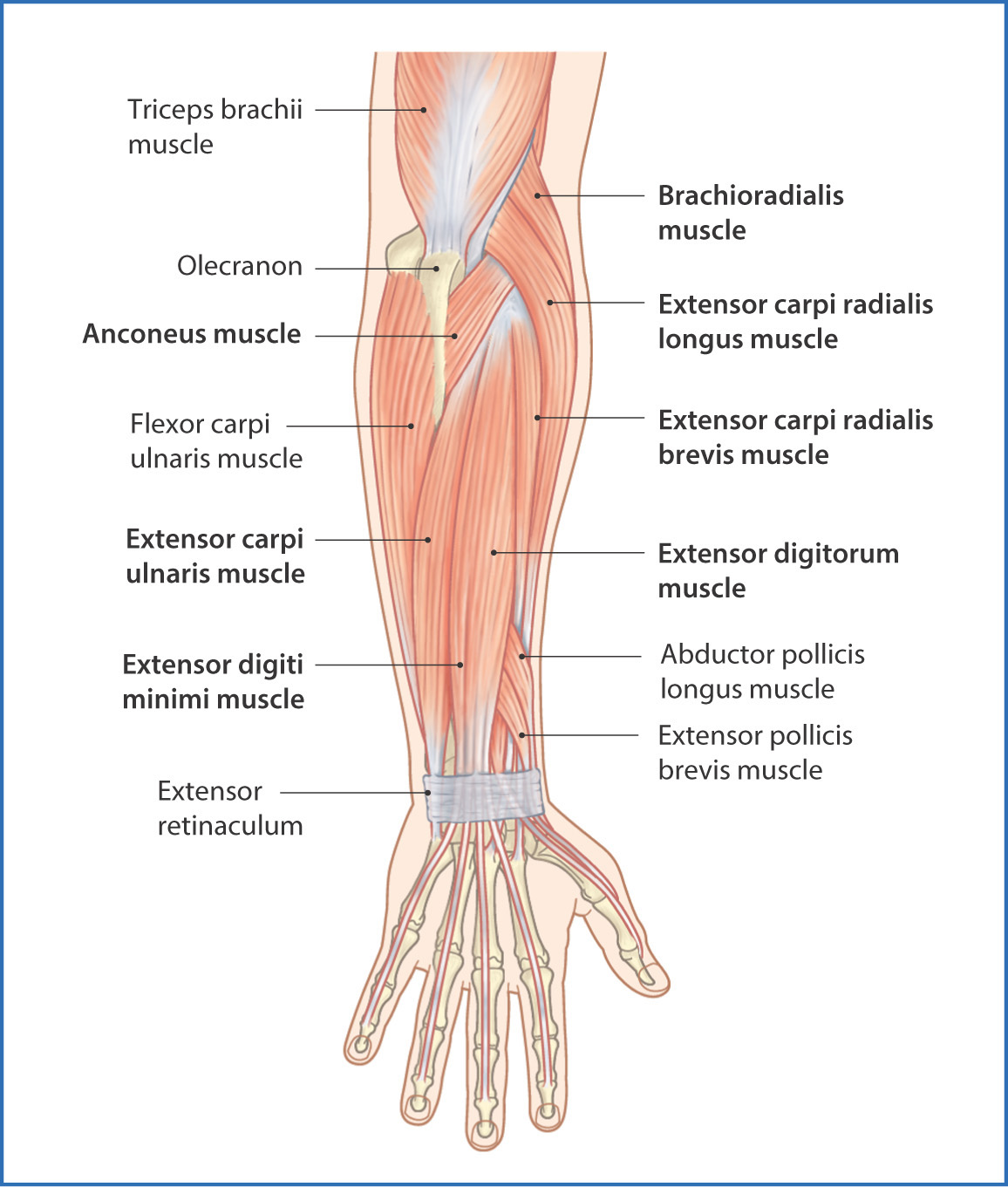
The posterior compartment of the forearm (or extensor compartment) contains twelve muscles which are chiefly responsible for extension of the wrist and digits, and supination of the forearm. Human anatomy for the artist the dorsal forearm part 2. Select from premium tendon stock of the highest quality. Muscles and tendons of the arm art print poster medical. Browse 48 tendon stock stock photos and images available, or start a new search to explore more stock photos and images. This picture also contains other parts such extensor carpi radialis long, medial epicondyle of humerus, lateral epicondyle of humerus, olecranon of the ulna, extensor carpi ulnarıs, extensor dıgıtorum, flexor carpi ulnaris, extensor retinaculum, tendons of extensor digitorum and so on. Tendons and ligaments are bands of connective tissue that help stabilize the body and allow movement. 397 x 283 jpeg 31kb. It is characterized by inflammation of the common extensor tendon where it attaches to the humerus, often as a result of tiny tears in the tendon that are not given sufficient time to heal, and is felt as pain that can radiate all the. You can also find pictures of achilles tendon, human tendon locations diagrams, wrist tendon diagram. One tendons inserts onto the forearm bone, the radius, and the second spreads out to join the fascia along the upper part of the forearm. Resting the muscles in the affected tendons is crucial to treating appreciated the pictures with written instructions. Tendons are the connective tissues that connect muscle to bone.