Upper Thigh Muscle Anatomy Mri - The Knee Mri Atlas Of Anatomy In Medical Imagery - Anterior superior iliac spine insertion:
byAdmin•
0
Upper Thigh Muscle Anatomy Mri - The Knee Mri Atlas Of Anatomy In Medical Imagery - Anterior superior iliac spine insertion:. Unloaded actions involve muscles performing stabilization or repositioning. The thigh has some of the body's largest muscles. Thigh muscles are responsible for allowing normal gait and proper lower extremity function(1). .anatomy mri, thigh muscle anatomy radiology, thigh muscles anatomy youtube, human muscles, thigh muscle anatomy cross sectional, thigh muscle of shoulder joint, muscle anatomy shoulder back, muscle anatomy shoulder upper arm, human muscles, muscle anatomy neck and shoulder. It is part of the lower limb.
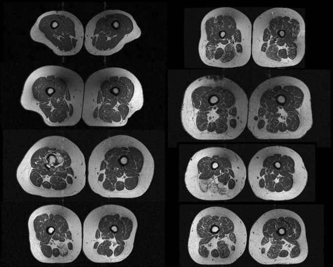
Mri patterns of neuromuscular disease involvement thigh & other muscles 2. .anatomy mri, thigh muscle anatomy radiology, thigh muscles anatomy youtube, human muscles, thigh muscle anatomy cross sectional, thigh muscle of shoulder joint, muscle anatomy shoulder back, muscle anatomy shoulder upper arm, human muscles, muscle anatomy neck and shoulder. Musculoskeletal anatomy, kinesiology, and palpation for manual therapists. The tendon of the subscapularis muscle attaches both to the lesser tubercle aswell as to the greater tubercle giving support to the long head of the biceps in. Robin smithuis and henk jan van der woude.
Clinical Evaluation Of Fully Automated Thigh Muscle And Adipose Tissue Segmentation Using A U Net Deep Learning Architecture In Context Of Osteoarthritic Knee Pain Springerlink from media.springernature.com Learn about the anatomy of the hamstrings, the group of muscles at the back of the upper leg, plus table of contents. Both supinator and pronator teres muscles have their origins on the humerus and ulna and insert on opposite sides of the radius to roll the wrist in opposite directions. Both the thigh and leg are divided into three separate compartments. Anatomy of the thigh : • acromion • clavicle • deltoid ( im injections) • humerus • biceps muscle • biciptal groove • brachila pulse( blood b) supplies most of the intrinsic muscles of the hand including the hypothenar eminence, and skin on the medial side of the hand. Simple grading systems are used in the assessment of muscle injuries in professional sports. Muscle mri can provide information that is complementary to clinical, histologic, genetic, and laboratory findings for the diagnosis of neuromuscular disease. The thigh is the area between the hip and the knee joint.
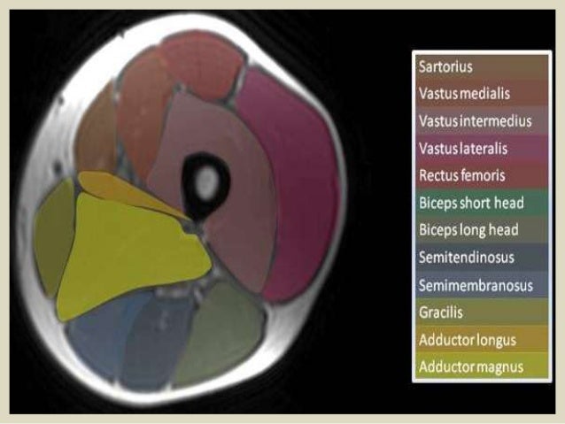
A magnetic resonance imaging (mri) was performed on a healthy subject;
The thigh is the area between the hip and the knee joint. The uppermost of the medial thigh muscles is the pectineus muscle. Both supinator and pronator teres muscles have their origins on the humerus and ulna and insert on opposite sides of the radius to roll the wrist in opposite directions. As the name implies they adduct the thigh at the hip. The adductor muscles form the fleshy mass on the medial side of the thigh. The gold standard for diagnosis of this condition is electromyography. Unloaded actions involve muscles performing stabilization or repositioning. The anterior femoral muscles (fig. It arises by tendinous fibers from the anterior superior iliac spine and the upper half of the notch below it. Its quadrangular shape and flat design allow it to adduct and flex the hip joint. Anatomy of the thigh : A magnetic resonance imaging (mri) was performed on a healthy subject; This is a table of skeletal muscles of the human anatomy.
Mri gives a detailed view of muscle injury. The uppermost of the medial thigh muscles is the pectineus muscle. Choose from 500 different sets of flashcards about thigh muscle anatomy on quizlet. Upper medial surface of the shaft of the tibia in front of the insertions of the gracilis and the semitendinosus nerve supply: Both the thigh and leg are divided into three separate compartments.
The Hip Anatomy On 3t Mr And 3d Pictures from www.imaios.com There are around 650 skeletal muscles within the typical human body. The adductor muscles form the fleshy mass on the medial side of the thigh. Muscles in the posterior compartment of the thigh. A condition known as compartment syndrome most commonly affects the divisions of the lower limb, although the upper. Typical findings are edema, hematoma, and partial or complete muscles tears. The thigh is the area between the hip and the knee joint. Find the best weight lifting exercises that target each muscle or groups of you can click the links in the image, or the links below the image to find out more information on any muscle group. Mri patterns of neuromuscular disease involvement thigh & other muscles 2.
There are around 650 skeletal muscles within the typical human body.
It is part of the lower limb. Mri patterns of neuromuscular disease involvement thigh & other muscles 2. Muscle mri allows the identification of edema and fatty replacement of muscle tissue. Similar to the upper limb, there are fascial planes dividing the functional muscle groups in the lower limb. Similar to fkrp distinguishing feature obturator externus & internus less involved than fkrp upper body common: Typical findings are edema, hematoma, and partial or complete muscles tears. Dummies has always stood for taking on complex concepts and making them easy to understand. Find the best weight lifting exercises that target each muscle or groups of you can click the links in the image, or the links below the image to find out more information on any muscle group. Learn about thigh muscle anatomy with free interactive flashcards. Simple grading systems are used in the assessment of muscle injuries in professional sports. Upper medial surface of the shaft of the tibia in front of the insertions of the gracilis and the semitendinosus nerve supply: Almost all muscles cross at least one joint (moveable connection between two bones) and cause an action across that joint. The gold standard for diagnosis of this condition is electromyography.
Dummies helps everyone be more knowledgeable and confident in applying what they know. Whether it's to pass that big test, qualify for that big promotion or even master that cooking technique; 15 17 magnetic resonance imaging (mri): The uppermost of the medial thigh muscles is the pectineus muscle. It arises by tendinous fibers from the anterior superior iliac spine and the upper half of the notch below it.
Presentation1 Pptx Radiological Anatomy Of The Thigh And Leg from image.slidesharecdn.com There are around 650 skeletal muscles within the typical human body. Anatomy of the thigh : Hand anatomy yoga anatomy anatomy study anatomy reference wrist anatomy upper limb anatomy medical anatomy human anatomy and physiology medical coding. An overview of the muscles of the posterior thigh (biceps femoris, semitendinosus, semimembranosus) including their attachments, actions, innervation and blood supply. Dummies has always stood for taking on complex concepts and making them easy to understand. The gold standard for diagnosis of this condition is electromyography. Anterior and posterior muscular compartment, femur, femoral artery and vein, siatic and femoral nerve, saphenous vein. A magnetic resonance imaging (mri) was performed on a healthy subject;
Anterior and posterior muscular compartment, femur, femoral artery and vein, siatic and femoral nerve, saphenous vein.
15 17 magnetic resonance imaging (mri): Learn about thigh muscle anatomy with free interactive flashcards. The muscles of the torso, examined in the previous chapter, include a few that attach directly into the upper arm and help move the humerus at the shoulder joint. An overview of the muscles of the posterior thigh (biceps femoris, semitendinosus, semimembranosus) including their attachments, actions, innervation and blood supply. Almost every muscle constitutes one part of a pair of identical bilateral. Robin smithuis and henk jan van der woude. Typical findings are edema, hematoma, and partial or complete muscles tears. Dummies has always stood for taking on complex concepts and making them easy to understand. .anatomy mri, thigh muscle anatomy radiology, thigh muscles anatomy youtube, human muscles, thigh muscle anatomy cross sectional, thigh muscle of shoulder joint, muscle anatomy shoulder back, muscle anatomy shoulder upper arm, human muscles, muscle anatomy neck and shoulder. Anatomy of the human body. Magnetic resonance imaging (mri) can be beneficial in identifying adductor brevis or adductor longus muscle atrophy which would indicate possible obturator nerve entrapment. The gold standard for diagnosis of this condition is electromyography. There are several ways to do this.
The thigh is the area between the hip and the knee joint upper thigh anatomy. Almost all muscles cross at least one joint (moveable connection between two bones) and cause an action across that joint.